Un-Complicating the Complicated: Part 2 Fascial Lines
Alrighty, let’s get started with Part 2.
While I initially planned on only having two parts to this series there is going to be more…
The reason there is going to be more is because fascia is spectacular and fascia is one of the major role players in my practice. I practice QSM3 Chiropractic which looks at the compensation patterns of the musculoskeletal and fascial system and then based on the pattern, certain adjustments are put on specific areas in the lower back, hips, upper back and neck in order to reduce the tension along the fascial lines and help return the body to an upright symmetrical position. Add onto that proprioceptive rehabilitation and neuromuscular rehabilitation and you get some pretty spectacular results and resolution of many painful syndromes.
Basically all that to say I really like fascia and this is the perfect time for me to explain it in ever more detail than I initially planned.
Last week we ended with discussing the different fascial lines that connect different groups of muscles. Today we are going to dive deeper into those fascial lines, discuss certain connections, the muscles involved and some typical pain syndromes that show up along these fascial lines.
This brings us back to the Superficial Back Line.
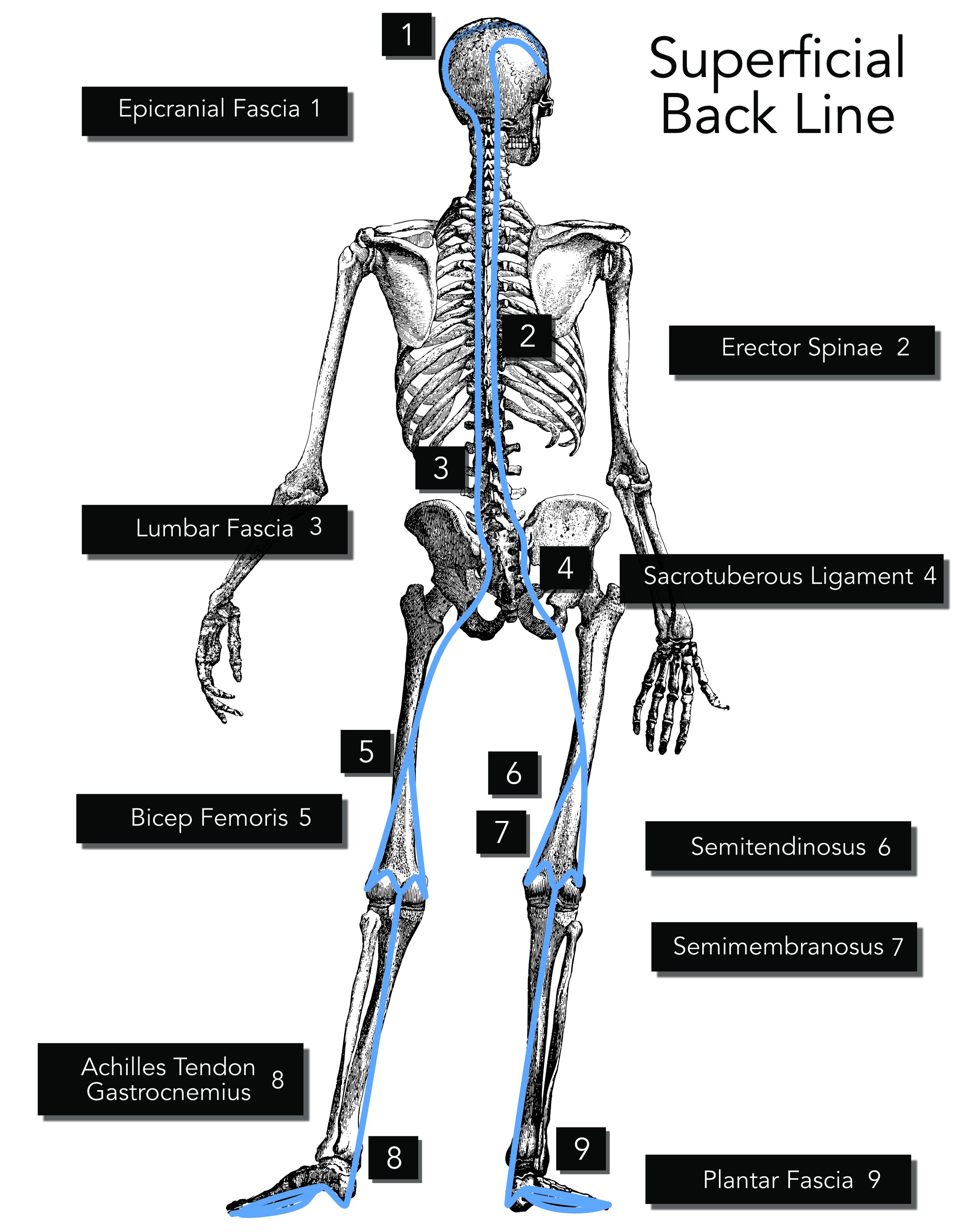
The Superficial Back Line (SBL) is a continuous line of fascia and muscle that runs along the backside of the body. It plays a critical role in maintaining posture and facilitating movements, such as standing, walking, and bending backward and forward.
The SBL literally runs from your foot all the way to your skull. It starts at the plantar fascia(an area at the bottom of our foot) goes up around our achilles tendon, along the calf(gastrocnemius), along the hamstrings, connects to the pelvis and the sacrum, then continues up the back muscles(specifically the erector spinae), then up to the back of the neck to the skull connecting to the epicranial fascia. A further explanation of the different areas the SBL connects with is below.
Plantar fascia: The plantar fascia is a thick, fibrous band of connective tissue on the bottom of the foot. It connects the heel bone to the toes and supports the foot's arch, helping to absorb and distribute forces during walking and running.
Achilles tendon: The Achilles tendon is the largest and strongest tendon in the body. It connects the calf muscles (gastrocnemius and soleus) to the heel bone (calcaneus) and enables the foot to push off the ground during walking, running, and jumping.
Gastrocnemius: The gastrocnemius is the most superficial muscle of the calf, forming the bulge on the back of the lower leg. It is involved in plantarflexion of the foot, knee flexion, and plays a role in maintaining an upright posture.
Hamstrings: The hamstrings are a group of three muscles (semitendinosus, semimembranosus, and biceps femoris) located at the back of the thigh. They are responsible for flexing the knee, extending the hip, and helping to maintain the pelvis in an upright position.
Sacrotuberous ligament: The sacrotuberous ligament is a strong, fibrous band that connects the sacrum (lower part of the spine) to the ischial tuberosity (sit bone). It provides stability to the pelvis and transfers forces between the spine and lower limbs.
Erector spinae: The erector spinae is a group of muscles and tendons that run along the length of the spine. These muscles extend the vertebral column, maintaining an upright posture, and provide side-to-side stability.
Occipitalis: The occipitalis is a thin, flat muscle located at the back of the skull. It connects to the galea aponeurotica (a fibrous tissue layer) and assists in raising the eyebrows and moving the scalp.
As you can see the SBL's interconnected nature means that it is involved in a lot and dysfunction in one part of the line can lead to compensations and imbalances in other parts, potentially causing pain and movement limitations. It is essential to recognize that the interconnected nature of the SBL means that dysfunction or pain in one area can contribute to or exacerbate issues in other areas along the line. Some of the common issues that can be exacerbated of caused by dysfunction or injury to the SBL include:
Plantar fasciitis: Pain and inflammation in the plantar fascia, a thick band of tissue running along the sole of the foot, connecting the heel to the toes.
Achilles tendinopathy: Pain, swelling, and stiffness in the Achilles tendon, which connects the calf muscles to the heel bone.
Hamstring strains or injuries: Overstretching, tearing, or inflammation of the hamstring muscles located at the back of the thigh.
Sacroiliac joint dysfunction: Pain and discomfort in the sacroiliac joint, which connects the sacrum to the ilium in the pelvis, often caused by excessive or reduced motion in the joint.
Lower back pain: Pain or discomfort in the lumbar region, which can have various causes, such as muscle strains, ligament sprains, disc herniation, or facet joint dysfunction.
Thoracic outlet syndrome: Compression of nerves and blood vessels between the collarbone and first rib, causing pain in the neck and shoulder, and numbness or tingling in the fingers.
Tension headaches: Pain or discomfort, often described as a tight band around the head, caused by muscle tension or stress, which can involve the occipitalis muscle at the base of the skull.
Now it is important to note that, just because you have lower back pain it doesn’t necessarily mean it is directly related to the SBL. Same with plantar fasciitis, hamstring injury, or any of the other issues above. It does mean that it can be a contributing factor or even a cause in some cases, which is why a thorough examination is key when working on resolving these issues.
Next is the Superficial Front Line.
The Superficial Front Line (SFL) is an interconnected chain of muscles and fascia that runs along the anterior (front) aspect of the body, from the top of the feet to the sternocleidomastoid and then to the back of the skull.
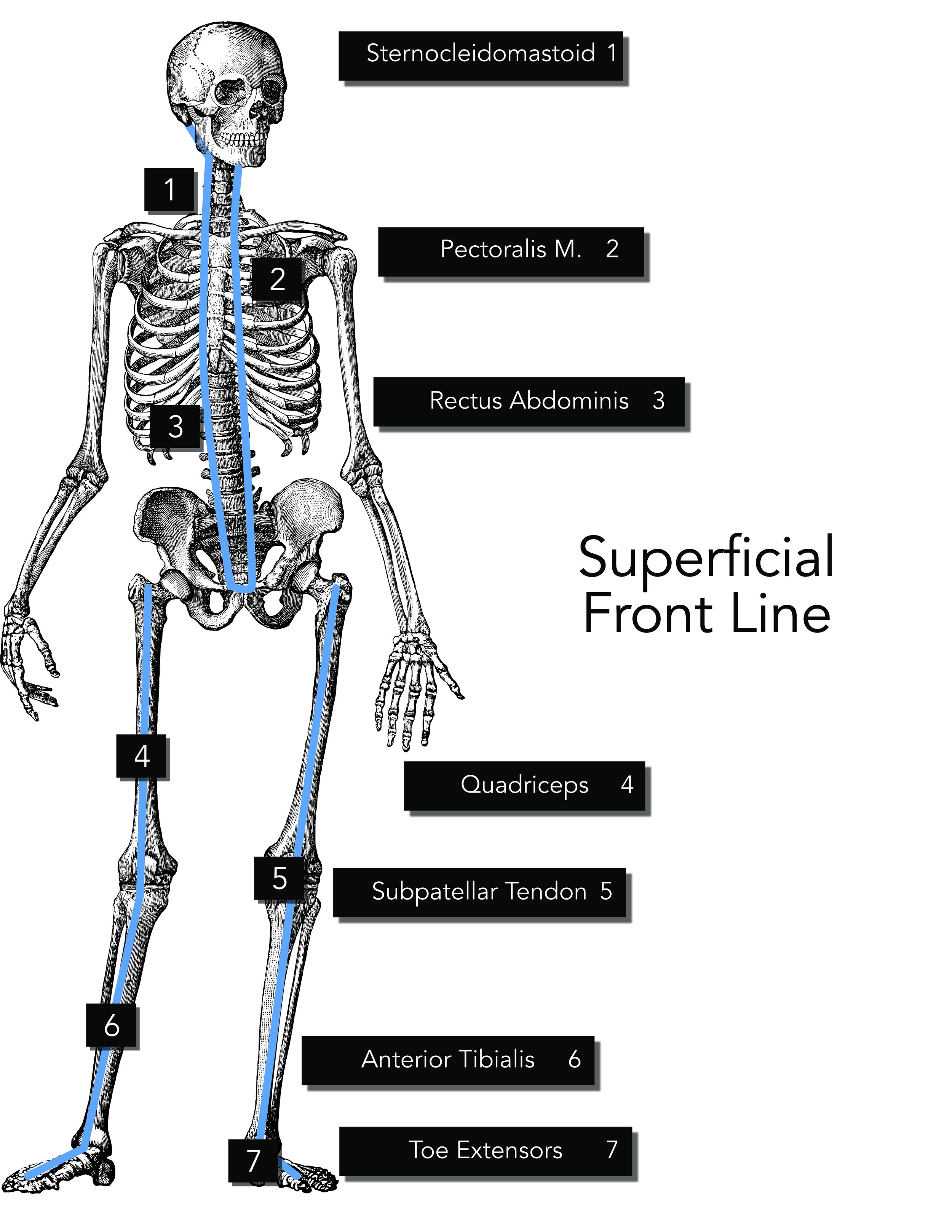
This myofascial line plays a crucial role in maintaining an upright posture, stabilizing the body during movement, and facilitating flexion movements. Let's explore the key structures and their functions within the SFL:
Tibialis anterior: This muscle, located at the front of the lower leg (shin), helps dorsiflex and invert the foot, providing stability during walking and running.
Extensor digitorum longus: This muscle, also in the lower leg, is responsible for extending the toes and assisting in dorsiflexing the foot.
Rectus femoris: As part of the quadriceps muscle group, the rectus femoris is vital for knee extension and hip flexion, allowing for movements such as walking, running, and jumping.
Quadriceps: A group of four muscles, including the rectus femoris, that work together to extend the knee and stabilize the leg during movement.
Rectus abdominis: Commonly referred to as the "abs," this muscle runs vertically along the front of the abdomen, playing a significant role in trunk flexion, maintaining posture, and providing core stability.
Pectoralis major: The large chest muscle responsible for shoulder flexion, adduction, and medial rotation, allowing for movements like pushing and lifting.
Sternocleidomastoid: This muscle runs diagonally from the sternum and clavicle to the mastoid process behind the ear, helping to flex and rotate the head and neck.
The SFL is similar to the SBL in its interconnected nature means that dysfunction, restriction, or imbalance in one area can impact other areas within the line, potentially causing pain or movement limitations.
Some issues that can arise when there are injuries along the SFL include,
Shin splints: Pain along the tibia (shinbone) caused by inflammation of the muscles, tendons, and connective tissues, often associated with the tibialis anterior muscle.
Patellar tendinitis: Inflammation of the patellar tendon connecting the kneecap (patella) to the shinbone, often due to overuse or excessive strain on the quadriceps muscles.
Iliopsoas syndrome: Pain and inflammation in the hip flexor muscles (iliopsoas), which can lead to lower back and hip pain, often resulting from muscle imbalances, excessive tension, or overuse.
Rectus abdominis strain: Strain or tear in the rectus abdominis muscle, which can cause pain in the abdomen, often due to overuse or sudden, forceful contraction during activities like sit-ups or twists.
Anterior neck pain: Pain in the front of the neck, which can result from tension or strain in the muscles and fascial structures of the anterior neck, including the sternocleidomastoid and scalene muscles.
This brings us to the last line we are going to be talking about today which is the Lateral Line.
The Lateral Line also has connections to the Sternocleidomastoid along with the Splenius Capitis and it is a continuous line of fascia and muscles running along the sides of the body, connecting structures from the head to the feet. This line plays an important role in stabilizing the body during side-to-side movements, providing lateral support, and contributing to maintaining balance.
Let's explore the key structures and their functions within the LL:
Peroneus muscles (Peroneus longus and brevis): Located on the lateral side of the lower leg, these muscles help stabilize the ankle and control eversion of the foot.
Iliotibial (IT) band: A thick, fibrous band of fascia that runs along the lateral thigh, connecting the tensor fasciae latae and gluteus maximus muscles to the tibia.
Tensor fasciae latae (TFL): A small muscle located at the front of the hip, which helps stabilize the pelvis and assists in hip abduction and flexion.
Gluteus medius and minimus: Muscles situated on the lateral side of the hip, responsible for hip abduction and rotation.
Quadratus lumborum: A deep muscle in the lower back that connects the pelvis to the spine, playing a crucial role in stabilizing the lumbar spine and assisting in lateral flexion of the trunk.
External and internal obliques: Abdominal muscles that contribute to the rotation and lateral flexion of the trunk.
Internal and External Intercostal Muscles: These muscles are found between the ribs and play a significant role in respiration. The external intercostals are involved in inspiration (inhaling), while the internal intercostals are involved in forced expiration (exhaling forcefully).
Those are some of the different fascial line and they play a key role in our movement and stability. In this next section we’re going to discuss how these lines transmit force and introduce the concept of tensegrity.
Transmitting Force
The Superficial Front Line (SFL), Superficial Back Line (SBL), and Lateral Line are integral to force transmission in the body. These interconnected chains of muscles and fascia work together to distribute and transmit forces during various movements and activities, contributing to overall biomechanics and efficiency.
Superficial Front Line (SFL): The SFL connects muscles along the body's anterior chain, from the top of the feet to the neck. When these muscles contract in a coordinated manner, they create tension through the entire line, helping to transmit forces during flexion movements such as walking, running, or jumping. For example, during the push-off phase of walking or running, the SFL muscles help transmit force from the feet through the lower and upper limbs, and into the trunk, promoting efficient movement and reducing the risk of injury.
Superficial Back Line (SBL): The SBL runs along the body's posterior chain, connecting muscles from the feet to the head. It plays a significant role in maintaining an upright posture and facilitating extension movements. When the muscles in the SBL contract, they generate tension throughout the entire line, enabling force transmission during extension movements such as standing up, walking, or running. For instance, during the stance phase of walking or running, the SBL muscles transmit forces from the ground up through the lower limbs, trunk, and into the head, providing stability and support to the entire body.
Lateral Line: The Lateral Line connects muscles along the sides of the body, from the head to the feet. This line is responsible for stabilizing the body during side-to-side movements and maintaining balance during various activities. The interconnected muscles and fascia in the Lateral Line transmit forces during lateral movements, such as side-stepping or changing direction while running. For example, when changing direction quickly, the Lateral Line muscles on one side of the body contract and generate tension through the entire line, allowing for efficient force transmission and helping to maintain balance and stability.
These three lines work together to transmit forces efficiently and maintain stability during a wide range of movements. By distributing forces across multiple muscles and joints, the body can effectively minimize the risk of injury and enhance overall biomechanical efficiency.
But how??
Okay, so these different fascial lines work to transmit force, but how does that work?
When we typically think about a muscle transmitting force, we often view it through a reductionist lens. For instance, if you think of doing a bicep curl, we tend to believe it is the bicep doing all the work. However, this perspective, largely influenced by Newtonian biomechanics, oversimplifies the complex process of human movement. Newtonian biomechanics focuses on linear forces, mechanical levers, and joints, and while it provides valuable insights into certain aspects of movement it doesn’t fully explain how our bodies can efficiently perform a wide range of motions. (I expand on this in a section at the conclusion of this blog)
Swanson RL 2nd. Biotensegrity: a unifying theory of biological architecture with applications to osteopathic practice, education, and research--a review and analysis. PMID: 23329804.
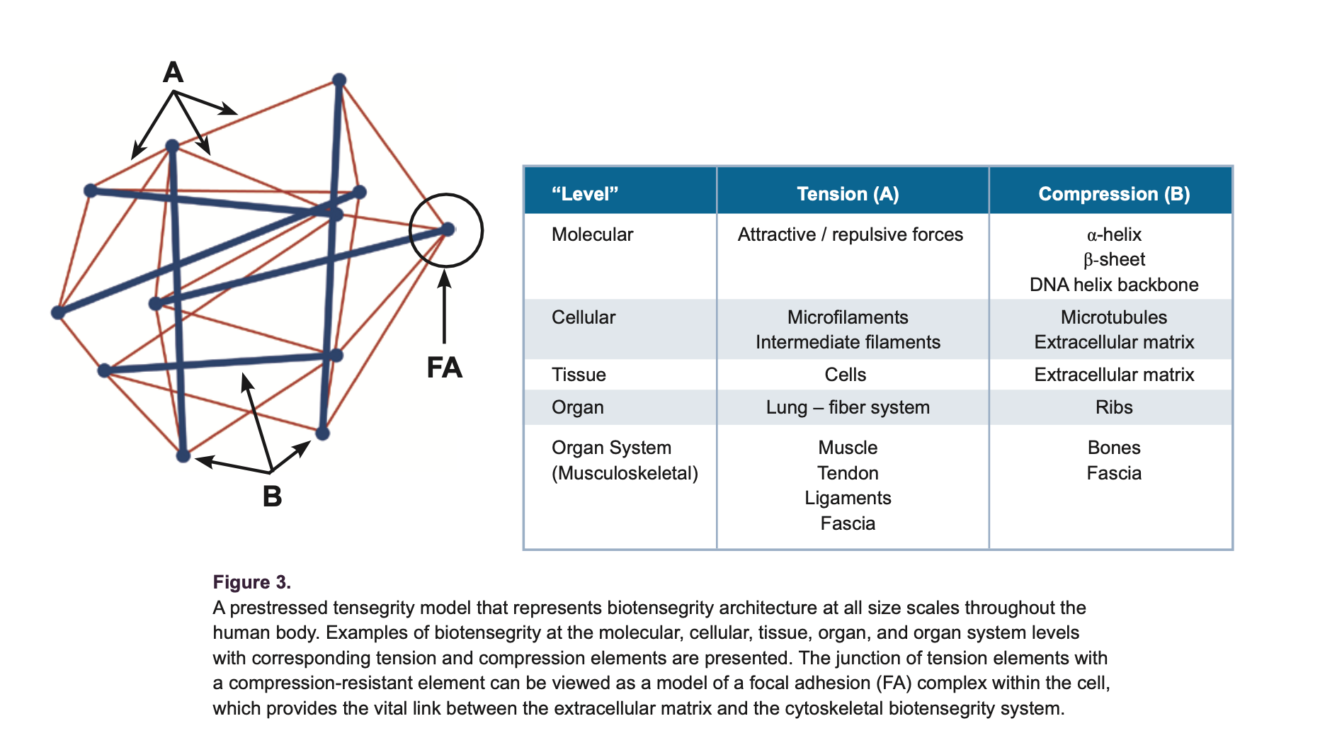
This is where the concept of tensegrity, or tensional integrity, comes into play. Tensegrity is a structural principle based on the balance between tension and compression components, where continuous tension members (such as fascia) and discontinuous compression members (such as bones) maintain a stable structure. In the human body, tensegrity is essential for maintaining stability and facilitating efficient movement. Rather than isolating specific muscles or joints, tensegrity highlights the interdependence and interconnectedness of all body structures.
Fascia plays a crucial role in the tensegrity model of the human body. It forms a continuous network that connects muscles, bones, and organs, allowing for efficient force transmission and distribution across multiple structures.
When performing a bicep curl, for example, it's not just the bicep muscle working in isolation. Instead, the fascial network, through the principles of tensegrity, allows other parts of the body to assist in lifting the weight. This interconnectedness helps to distribute forces, reduce the risk of injury, and optimize movement efficiency.
How we move goes beyond the traditional Newtonian biomechanics perspective. Understanding the concept of tensegrity and the role of fascia in the body provide a more comprehensive understanding of how our bodies can efficiently move through the world.
Part 2 done! We will continue exploring fascia and the role it plays in our musculoskeletal health next week. I hope you enjoyed this blog post. Next week we will continue exploring the other fascial line as well as discuss the role these fascial line play in body alignment. Also below I dive a little deeper into Newtonian Physics and how it can’t fully explain how we move.
More on Tensegrity & Newtonian Physics
Let’s discuss the traditional approach to biomechanics, based on Newtonian physics. Only utilizing Newtonian physics simplifies the body's motion into linear and rotational movements, using equations that describe force, mass, and acceleration. While this approach has been useful in understanding basic mechanical principles, it falls short when trying to capture the complex, interconnected nature of the human body, especially when considering movement. Let’s look at this in relationship to the bicep curl like we discussed above.
There are several reasons why applying only Newtonian physics to the bicep curl might not fully capture the dynamics of the movement:
Non-linear properties of soft tissues: Newtonian physics assumes linear relationships between force and displacement. However, soft tissues like muscles, tendons, and fascia exhibit non-linear properties, meaning their response to force may not follow a simple linear pattern. This complicates the application of Newtonian mechanics to the bicep curl.
Interconnectedness: Newtonian physics often treats the body as a collection of separate segments or levers, with forces acting in isolation. In reality, the human body is a highly interconnected system, with forces being transmitted across multiple structures, such as fascial lines. This interconnectivity makes it difficult to analyze the bicep curl using only Newtonian principles.
Tensegrity: The concept of tensegrity highlights the importance of the continuous tension within the fascial network that maintains the body's structural integrity. This principle is not easily explained by Newtonian physics, which focuses on individual forces and isolated components rather than the integration of tension and compression elements throughout the body.
Neuromuscular control: Newtonian physics doesn't account for the complex neuromuscular control that regulates muscle activation, coordination, and timing during movement. The bicep curl involves the interaction of numerous muscles, nerves, and other soft tissues, making it challenging to analyze using only Newtonian mechanics.
While Newtonian physics has been useful in understanding some aspects of biomechanics, its limitations become apparent when trying to fully explain complex movements. Considering the interconnected nature of the human body and the non-linear properties of soft tissues, tensegrity and the fascial system and their interconnectedness better explains the dynamics of human movement.